Advocacy
Stakeholders meet on “Identification and Detection of Entamoeba histolytica” was conducted at Sri Balaji Vidyapeeth Deemed-to-be-University, Pondicherry on July 21, 2019. This programme was of national importance since the amoebiasis is being reported from different parts of India because of poor socioeconomic conditions and sanitation levels. This meeting was conducted with an objective to frame the guidelines on the identification and detection of E. histolytica with reference to conventional diagnostic methods and molecular diagnosis targeting appropriate genes of E. histolytica. The recommendations of the panel were released as a declaration on the diagnosis of amoebiasis and were circulated to various administrative and scientific bodies in India as reference policy document on the diagnosis of amoebiasis.
“PONDICHERRY DECLARATION ON IDENTIFICATION AND DETECTION OF ENTAMOEBA HISTOLYTICA- 2019”
We, the panel members at this meeting have arrived at a decisive consensus regarding the laboratory diagnosis of Intestinal and Extra-Intestinal amoebiasis (Entamoeba histolytica), with particular reference to Microscopy, immunodiagnosis (antigen detection and antibody detection) and Molecular diagnosis. We submit with conviction that these recommendations are for the kind and immediate perusal as well as subsequent implementation by the Apex Council, viz., Department of Health Research (DHR) for the larger benefit of the community.
Diagnosis of Intestinal Amoebiasis
Microscopy
- Microscopic examination of stool samples is not very useful in the specific identification and diagnosis of Entamoeba histolytica infection but can be used for screening of Entamoeba histolytica-look alike non-pathogenic species (e.g. Entamoeba dispar, Entamoeba moshkovskii) which cannot be differentiated from histolytica on microscopy.
Immunodiagnosis
- Antibody based serological tests may be useful in the diagnosis of amoebiasis in developed countries since histolyticainfection is uncommon. Whereas in developing countries, infection due to E. histolytica remains endemic. This makes definite diagnosis of amoebiasis by antibody detection difficult because of the difficulty to corroborate the present from past infection. However, negative test may rule out the possibility of invasive amoebiasis.
- Antibody detection in serum: This is not very useful for diagnosis in areas where amoebiasis is endemic. Positive test cannot differentiate between current and past infection.
- Faecal antigen detection: Specific antigen detection tests are available like Enzyme Linked Immunosorbant Assay (ELISA) and Immunochromatographic rapid kits (ICT), using Lectin antigen for histolytica in faecal samples. These may be used for specific diagnosis of E. histolytica infections.
(Note: Faecal antigen detection tests may not give the same sensitivity in cases who are on anti-amoebic treatment).
Molecular diagnosis
- Conventional, Nested or Real Time PCR should be used for the confirmation of identification and detection of histolytica infection as these can differentiate between E. dispar and E. moshkovskii. PCR has a very high sensitivity and specificity. PCR assay targeting 18SrDNA with species specific probes are the most common and recommended test.
- Although conventional PCR assays has been increasingly used for detection and differentiation of Entamoebaspecies, the disadvantages it faced were time consumption when large-scale sample processing was required, cost, inability to produce quantitative results and false positive results due to carry-over contamination.
Diagnosis of Extra-intestinal Amoebiasis
Microscopy
- This has a very limited role in the diagnosis of extra-intestinal amoebiasis such as amoebic liver abscess, as parasites would not be visible in the majority of aspirated liver pus samples.
Immunodiagnosis
- Antibody detection in serum: This is not very useful for diagnosis in areas where amoebiasis is endemic. Positive test cannot differentiate between current and past infection. However, negative test may rule out the possibility of invasive amoebiasis.
- Antigen detection in serum and amoebic liver abscess pus: Among the antigen detection assays, ELISA has been the widely used to study amoebiasis. ELISA remains an important diagnostic tool in patients with invasive amoebiasis. Commercial antigen detection tests are available, but they do not have good sensitivity for detection of histolytica in liver abscess and are not recommended.
(Note: Antigen detection tests may not give the same sensitivity in patients who are on anti-amoebic treatment).
Molecular diagnosis
- Conventional, Nested or Real Time PCR should be used for the confirmation of diagnosis of histolytica infection. PCR has a very high sensitivity and specificity. PCR assay targeting 18SrDNA with species specific probes are the most common and recommended test.
- Accurate identification of pathogenic histolyticafrom non-pathogenic Entamoeba species is crucial in the management of patients and epidemiological study of amoebiasis outbreaks. Molecular-based techniques have been proven to be adequate to satisfy these needs and hence has emerged as the gold standard diagnostic tests in the current era.
GENERAL RECOMMENDATION
E. histolytica may be infecting only a fraction of the population based on the data available from various studies which were mainly based on microscopy and/or serology and detect Entamoeba species. As these could not differentiate between the pathogenic and non-pathogenic species, there is an urgent need to initiate accurate prevalence based study in different parts of the country for estimating true prevalence of E. histolytica. There is also an urgent need to introduce specific tests for accurate diagnosis of E. histolytica infections at the field level.
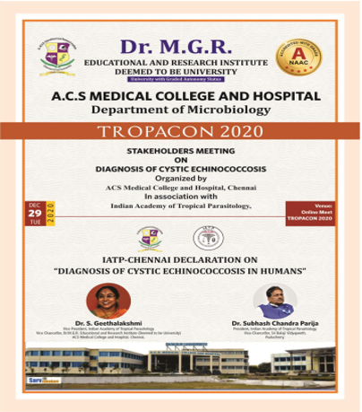
The panel members at this meeting have arrived at a consensus regarding the laboratory diagnosis of cystic echinococcosis, with particular reference to immunodiagnosis (antigen detection and antibody detection), microscopy and molecular diagnosis.

Stakeholders meet on “Identification and Detection of Entamoeba histolytica” was conducted at Sri Balaji Vidyapeeth Deemed-to-be-University, Pondicherry on July 21, 2019. This programme was of national importance since the amoebiasis is being reported from different parts of India because of poor socioeconomic conditions and sanitation levels. This meeting was conducted with an objective to frame the guidelines on the identification and detection of E. histolytica with reference to conventional diagnostic methods and molecular diagnosis targeting appropriate genes of E. histolytica. The recommendations of the panel were released as a declaration on the diagnosis of amoebiasis and were circulated to various administrative and scientific bodies in India as reference policy document on the diagnosis of amoebiasis.
“PONDICHERRY DECLARATION ON IDENTIFICATION AND DETECTION OF ENTAMOEBA HISTOLYTICA- 2019”
We, the panel members at this meeting have arrived at a decisive consensus regarding the laboratory diagnosis of Intestinal and Extra-Intestinal amoebiasis (Entamoeba histolytica), with particular reference to Microscopy, immunodiagnosis (antigen detection and antibody detection) and Molecular diagnosis. We submit with conviction that these recommendations are for the kind and immediate perusal as well as subsequent implementation by the Apex Council, viz., Department of Health Research (DHR) for the larger benefit of the community.
Diagnosis of Intestinal Amoebiasis
Microscopy
- Microscopic examination of stool samples is not very useful in the specific identification and diagnosis of Entamoeba histolytica infection but can be used for screening of Entamoeba histolytica-look alike non-pathogenic species (e.g. Entamoeba dispar, Entamoeba moshkovskii) which cannot be differentiated from histolytica on microscopy.
Immunodiagnosis
- Antibody based serological tests may be useful in the diagnosis of amoebiasis in developed countries since histolyticainfection is uncommon. Whereas in developing countries, infection due to E. histolytica remains endemic. This makes definite diagnosis of amoebiasis by antibody detection difficult because of the difficulty to corroborate the present from past infection. However, negative test may rule out the possibility of invasive amoebiasis.
- Antibody detection in serum: This is not very useful for diagnosis in areas where amoebiasis is endemic. Positive test cannot differentiate between current and past infection.
- Faecal antigen detection: Specific antigen detection tests are available like Enzyme Linked Immunosorbant Assay (ELISA) and Immunochromatographic rapid kits (ICT), using Lectin antigen for histolytica in faecal samples. These may be used for specific diagnosis of E. histolytica infections.
(Note: Faecal antigen detection tests may not give the same sensitivity in cases who are on anti-amoebic treatment).
Molecular diagnosis
- Conventional, Nested or Real Time PCR should be used for the confirmation of identification and detection of histolytica infection as these can differentiate between E. dispar and E. moshkovskii. PCR has a very high sensitivity and specificity. PCR assay targeting 18SrDNA with species specific probes are the most common and recommended test.
- Although conventional PCR assays has been increasingly used for detection and differentiation of Entamoebaspecies, the disadvantages it faced were time consumption when large-scale sample processing was required, cost, inability to produce quantitative results and false positive results due to carry-over contamination.
Diagnosis of Extra-intestinal Amoebiasis
Microscopy
- This has a very limited role in the diagnosis of extra-intestinal amoebiasis such as amoebic liver abscess, as parasites would not be visible in the majority of aspirated liver pus samples.
Immunodiagnosis
- Antibody detection in serum: This is not very useful for diagnosis in areas where amoebiasis is endemic. Positive test cannot differentiate between current and past infection. However, negative test may rule out the possibility of invasive amoebiasis.
- Antigen detection in serum and amoebic liver abscess pus: Among the antigen detection assays, ELISA has been the widely used to study amoebiasis. ELISA remains an important diagnostic tool in patients with invasive amoebiasis. Commercial antigen detection tests are available, but they do not have good sensitivity for detection of histolytica in liver abscess and are not recommended.
(Note: Antigen detection tests may not give the same sensitivity in patients who are on anti-amoebic treatment).
Molecular diagnosis
- Conventional, Nested or Real Time PCR should be used for the confirmation of diagnosis of histolytica infection. PCR has a very high sensitivity and specificity. PCR assay targeting 18SrDNA with species specific probes are the most common and recommended test.
- Accurate identification of pathogenic histolyticafrom non-pathogenic Entamoeba species is crucial in the management of patients and epidemiological study of amoebiasis outbreaks. Molecular-based techniques have been proven to be adequate to satisfy these needs and hence has emerged as the gold standard diagnostic tests in the current era.
GENERAL RECOMMENDATION
E. histolytica may be infecting only a fraction of the population based on the data available from various studies which were mainly based on microscopy and/or serology and detect Entamoeba species. As these could not differentiate between the pathogenic and non-pathogenic species, there is an urgent need to initiate accurate prevalence based study in different parts of the country for estimating true prevalence of E. histolytica. There is also an urgent need to introduce specific tests for accurate diagnosis of E. histolytica infections at the field level.
